Navigation |
The Most Common Types of Chronic Wounds and How They Are Treated Clinically
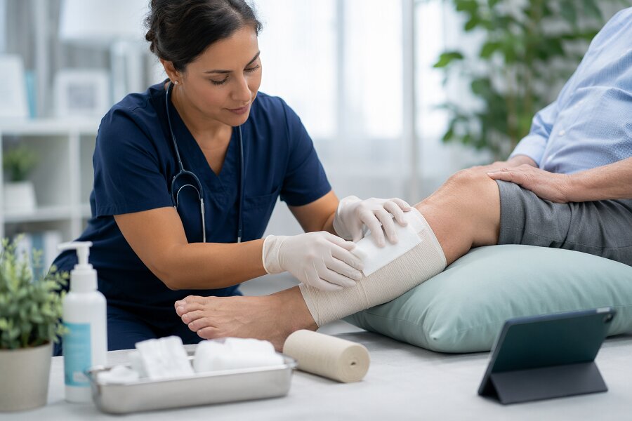
Each demands a different assessment, from vascular status to offloading needs, and a targeted plan. To choose the right approach, you first need to understand what type of wound you're dealing with and the underlying factors driving it. How Clinicians Define a Chronic WoundClinicians generally define a chronic wound as one that doesn't progress through the normal, orderly stages of healing within an expected period, usually around 4–6 weeks. This assessment relies on more than duration alone. Clinicians evaluate whether the wound is decreasing in size, forming healthy granulation tissue, and maintaining appropriate levels of exudate and inflammation. A wound that stops improving, deteriorates, or repeatedly breaks down after partial healing is typically classified as chronic. Documentation often includes wound depth, tissue types present, signs of local or systemic infection, and any history of recurrence, all of which support the determination that a wound is chronic. Why Some Wounds Don’t Heal: Key Risk FactorsHidden factors can interfere with wound healing, even when the injury appears simple. Healing depends on the body’s overall capacity to repair tissue, not just the condition of the skin surface. Reduced blood flow, diabetes, and smoking can limit the oxygen and nutrients reaching the wound. Ongoing pressure, friction, or persistent moisture can further damage the skin and delay closure. Additional risk factors include obesity, poor nutrition, and inadequate fluid intake, all of which can impair the body’s ability to rebuild tissue. Certain medications, such as long-term corticosteroids or chemotherapy agents, may slow the healing process by affecting inflammation or cell growth. Uncontrolled swelling (edema), infection, and poorly managed blood glucose can further disrupt normal repair mechanisms. Age-related changes, reduced mobility, and autoimmune conditions can also contribute, increasing the likelihood that an acute wound will become chronic. These factors all point to why wound care at home is rarely straightforward and why effective treatment must go beyond the wound itself to address the full picture of a patient's health. That is why professional mobile wound care services are essential, bringing clinical expertise, proper assessment tools, and evidence-based treatment directly to patients who need consistent, specialized care in the place they call home. The Main Types of Chronic Wounds in Clinical PracticeWhen these risk factors persist, certain wound types recur frequently in clinical practice and often fail to heal without targeted management. The most commonly encountered chronic wounds include pressure injuries, venous leg ulcers, arterial ulcers, mixed‑etiology leg ulcers, and non‑healing surgical or traumatic wounds. Pressure injuries typically develop over bony prominences when sustained pressure, shear, or friction exceed capillary perfusion pressure, leading to tissue ischemia and necrosis. Venous leg ulcers usually occur in the gaiter area of the lower leg and are often associated with chronic edema, lipodermatosclerosis, and hemosiderin staining, reflecting underlying venous hypertension. Arterial ulcers are more often located distally on the toes, feet, or lateral malleolus, and are usually painful, with dry, pale or necrotic wound beds and minimal exudate, consistent with impaired arterial perfusion. Mixed‑etiology ulcers show clinical features of both venous and arterial disease, and accurate diagnosis generally requires vascular assessment to guide treatment and avoid harm from inappropriate compression. Surgical and traumatic wounds become chronic when normal healing is disrupted by factors such as local ischemia, persistent infection or biofilm, excessive mechanical tension, repeated trauma, or systemic issues like poor glycemic control or malnutrition. In these cases, timely identification of the underlying cause and implementation of appropriate interventions are essential to promote progression through the normal phases of wound healing. Diabetic Foot Ulcers: Causes, Assessment, and TreatmentDiabetic foot ulcers (DFUs) result from a combination of peripheral neuropathy, peripheral arterial disease, and impaired immune function, making them a common and serious complication of diabetes. They usually develop over pressure points, areas of callus, or sites of minor trauma that may go unnoticed because of reduced sensation. Clinical assessment includes careful inspection of the skin, evaluation of ulcer size and depth (often by gentle probing), and assessment of vascular status through palpation of peripheral pulses, capillary refill, and measurement of the ankle–brachial index. Sensory testing is commonly performed with a 10‑g monofilament to detect loss of protective sensation. If infection is suspected, clinicians obtain wound cultures, and imaging is used to evaluate for underlying osteomyelitis or other bony involvement. Management centers on regular sharp debridement of nonviable tissue, appropriate antimicrobial therapy when infection is present, and use of moisture-balanced dressings to support wound healing. Offloading of pressure with specialized footwear, insoles, total contact casts, or other devices is essential to reduce mechanical stress on the ulcer. Optimizing glycemic control, discontinuing tobacco use, and maintaining regular follow‑up are important to reduce the risk of complications, including lower-extremity amputation. Venous Leg Ulcers: Compression and Core TherapiesVenous leg ulcers are a common and persistent type of chronic wound, most often resulting from long‑standing venous insufficiency in the lower limbs. They're typically associated with edema, aching, and shallow, exudative ulcers located in the gaiter region. Compression therapy is the central component of management. Common approaches include multilayer compression wraps, short‑stretch bandages, and medical‑grade compression stockings, all aimed at reducing venous hypertension and improving venous return. These measures are usually combined with leg elevation and strategies to enhance calf‑muscle pump function. Local wound care focuses on thorough wound bed preparation, which may involve debridement, use of moisture‑balancing dressings, and appropriate infection control. Additional aspects of care include managing associated dermatitis, addressing pain, optimizing nutritional status, and providing patient education to support correct and sustained use of compression over the long term. Arterial and Ischemic Ulcers: When Blood Flow FailsArterial and ischemic ulcers result from impaired arterial blood flow and therefore differ substantially from venous leg ulcers in both presentation and management. They typically occur on the toes, feet, or distal lower legs and often have well‑defined, “punched‑out” margins. The wound bed may appear pale, dry, or necrotic, and patients frequently report significant pain, which often increases when the limb is elevated due to further reduction in perfusion. Assessment should prioritize evaluation of arterial circulation. This usually includes ankle‑brachial index measurement, toe pressure assessment, and Doppler studies, with angiography considered when revascularization is being planned or when non‑invasive tests are inconclusive. Management focuses on restoring blood flow where possible, commonly through endovascular interventions or surgical bypass. In addition, strict control of cardiovascular risk factors, such as diabetes, hypertension, hyperlipidemia, and smoking cessation are essential. Local wound care is guided by the degree of ischemia and the risk of infection. In severely ischemic ulcers, maintaining a dry or only lightly moist wound environment and protecting the area from trauma are often recommended until perfusion is improved. High levels of compression are generally avoided in significant arterial disease, as they can further compromise blood flow; if mixed arterial‑venous disease is present, any compression must be carefully tailored based on objective vascular assessment. Pressure Injuries: Prevention, Offloading, and Bedside CarePressure injuries, unlike arterial and venous ulcers, result from sustained pressure, shear, or friction over bony prominences that compromises microcirculation and leads to tissue damage. Common locations include the heels, sacrum, hips, and ankles, particularly in patients who are immobile, critically ill, or have reduced sensory perception. Prevention focuses on minimizing the intensity and duration of pressure and shear forces, as well as maintaining skin integrity. Standard measures include repositioning patients at least every two hours (or more frequently based on risk and tolerance), using pressure-redistributing surfaces such as specialized mattresses or overlays, and elevating or “floating” the heels to remove direct pressure. Skin care involves keeping the skin clean, managing moisture from sweat, wound drainage, or incontinence, and using pH-balanced cleansers and moisturizers to reduce dryness and friction. Offloading is achieved with pillows, foam wedges, heel protectors, and appropriately fitted wheelchair cushions that distribute weight more evenly and reduce localized pressure. At the bedside, clinicians should perform regular skin inspections at a minimum once per shift, and more often in high-risk patients, with particular attention to bony prominences and any areas under medical devices. Additional preventive strategies include prompt incontinence management, use of barrier creams or moisture-associated skin damage (MASD) dressings in patients with frequent exposure to urine or stool, and protective dressings on high-risk areas to reduce friction and shear. Optimizing overall health (adequate protein and caloric intake, micronutrient support when indicated, and sufficient hydration) supports tissue perfusion and repair and reduces the risk of pressure injury development and delayed healing. Advanced Therapies and When to Refer or Consider SurgeryBeyond foundational wound care, some chronic wounds require advanced therapies or surgical intervention to achieve closure and preserve function. These options should be considered when a wound shows minimal or no improvement after 4–6 weeks of appropriate standard care, or when factors such as depth, infection, or ischemia increase the risk of tissue loss or systemic illness. Advanced therapies include negative pressure wound therapy, cellular or tissue-based products, topical growth factors, and hyperbaric oxygen therapy. These interventions are typically reserved for patients who've correctable underlying issues and are likely to benefit based on wound characteristics, comorbidities, and overall goals of care. Urgent referral for vascular assessment is indicated when peripheral pulses are diminished, the ankle–brachial index is reduced, or there are other signs of arterial insufficiency, as revascularization may be necessary for wound healing. Surgical consultation is important when there's exposed bone or tendon, suspected or confirmed deep infection (including osteomyelitis), unstable or function-limiting scarring, or concern for malignant transformation of a chronic wound (such as Marjolin ulcer). Early involvement of surgical and vascular specialists can improve diagnostic accuracy, guide appropriate intervention, and potentially reduce the risk of major amputation or other complications. ConclusionYou’ve seen how chronic wounds differ and why they don’t heal on their own. When you recognize the wound type and the underlying cause, you can choose targeted treatments like offloading, compression, pressure relief, or vascular workup that actually move healing forward. Don’t ignore stalled wounds; reassess early, involve specialists when needed, and use advanced therapies appropriately. Staying proactive and systematic gives every chronic wound the best chance to finally close.
|
User login |